2 Chromatic Transitions

Yellow, orange and red spread across the leaf in gradients, overlapping fields and fine, irregular markings. Colour becomes a way of approaching transformations that are only partly accessible through the visible surface. This chapter examines where colours meet, how they follow or cross the vein network, and how their distribution changes the appearance of the whole leaf. Microscopic views reveal textures and small transitions that recede within the larger image. Water loss, seasonal ageing and local changes in the tissue are considered as potentially overlapping processes whose individual contributions remain open to investigation. Together, the images form a chromatic record of leaf bodies in transition.
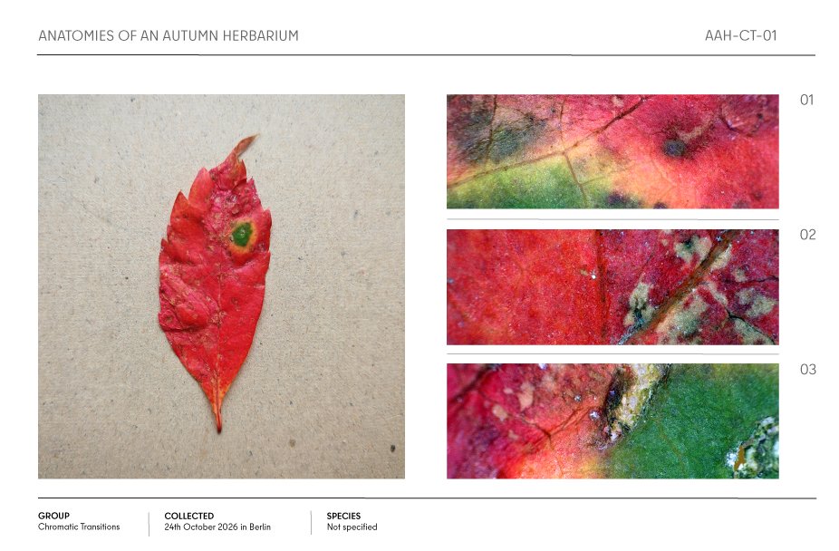
Red autumn leaf specimen with a green spot, shown alongside three colorful magnified detail views in an herbarium layout.

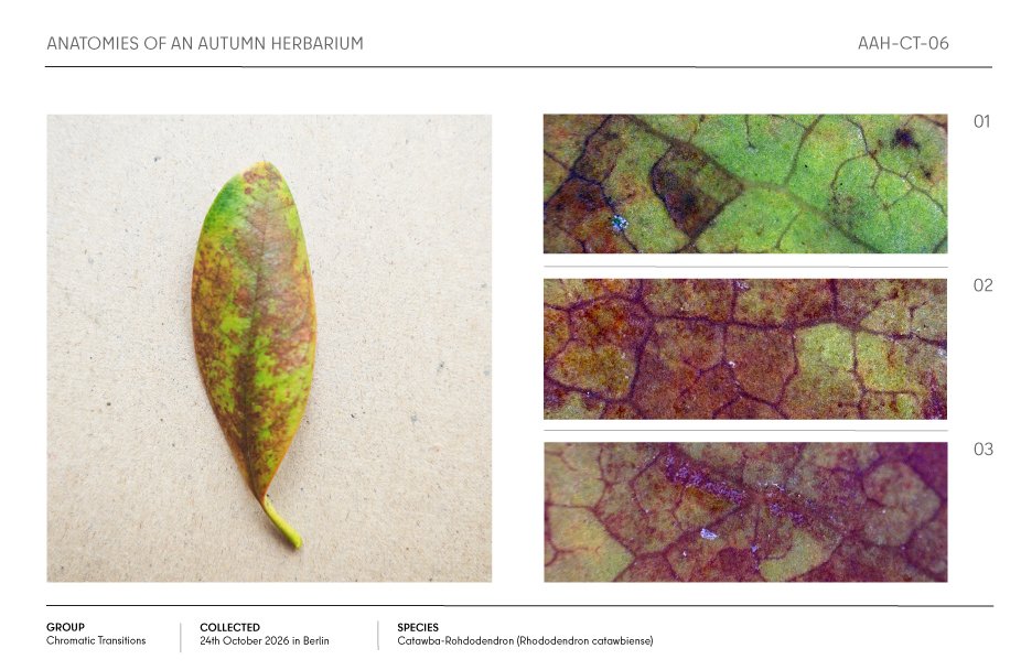
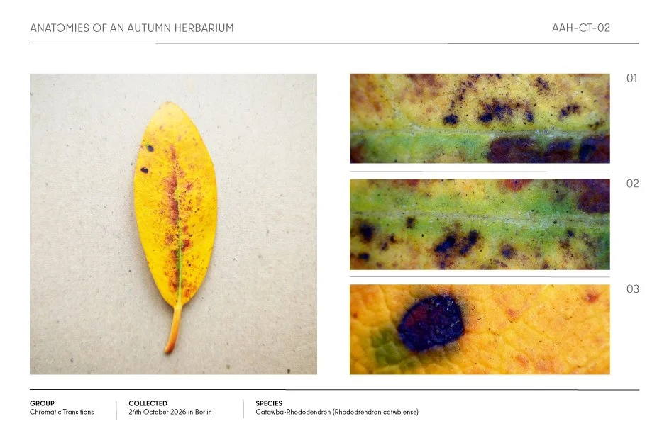
Autumn herbarium page featuring a yellow Catawba rhododendron leaf and three close-up images of its mottled surface.

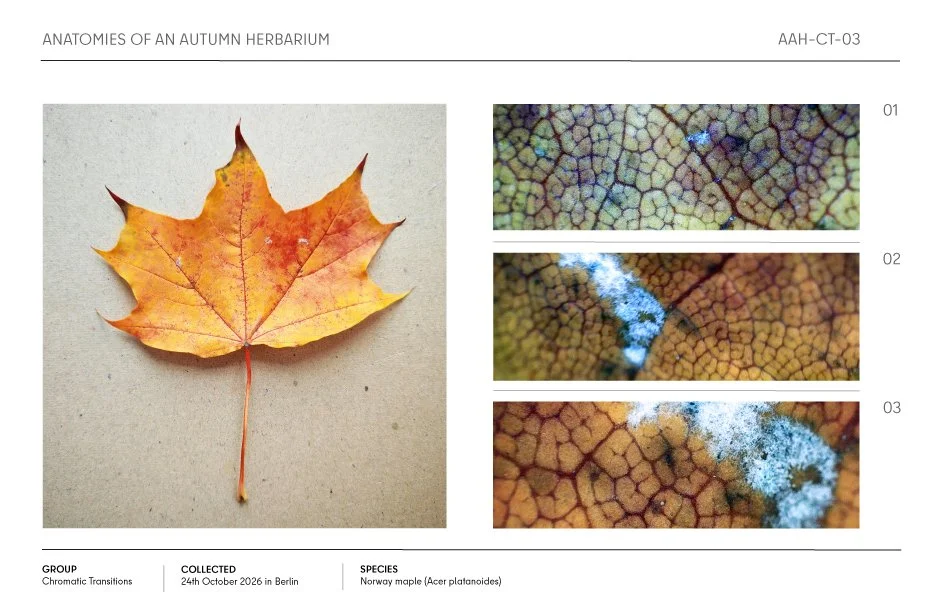
Autumn Norway maple leaf herbarium specimen with three microscope close-ups showing its changing cellular texture

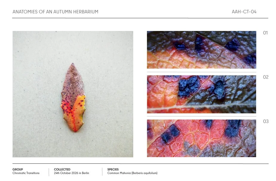
Autumn herbarium specimen of a colorful Mahonia (Berberis aquifolium) leaf with red, orange, purple, and blue chromatic transitions, shown with three close-up texture views.

Autumn Norway maple leaf herbarium specimen with orange-yellow foliage and close-up views of gray-blue mold growth

Autumn Catawba rhododendron leaf shown with three close-up views of its colorful vein and tissue patterns.

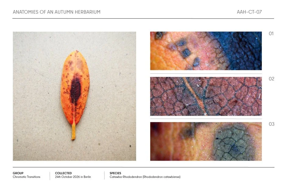
Autumn herbarium specimen sheet featuring an orange-red Catawba rhododendron leaf with three labeled close-up texture details.

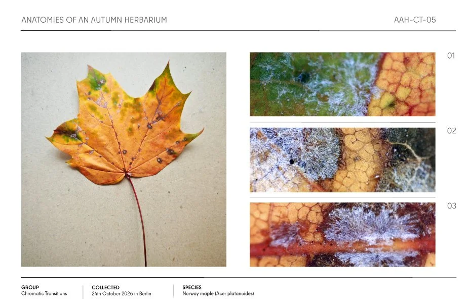
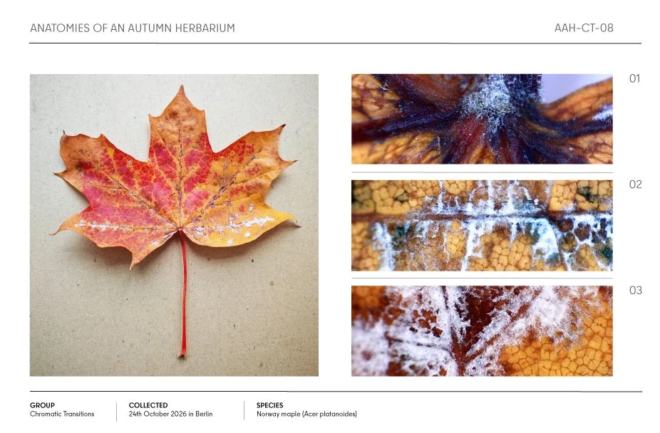
Autumn Norway maple leaf with red and yellow foliage, shown alongside three close-up views of white fungal growth.