3 Residual Green

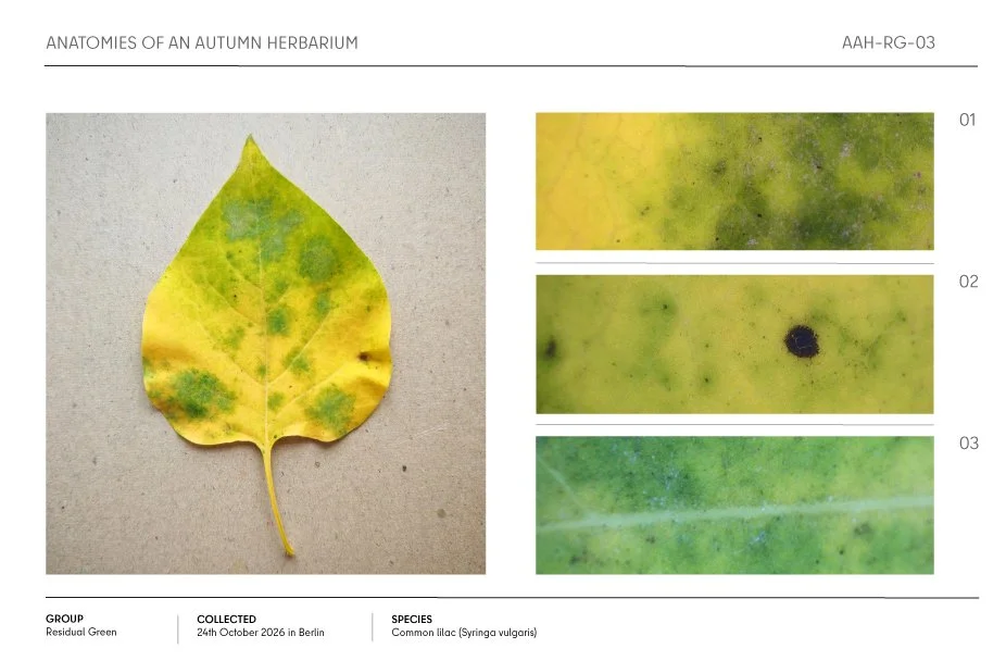
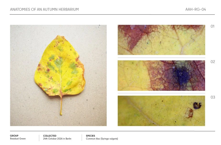
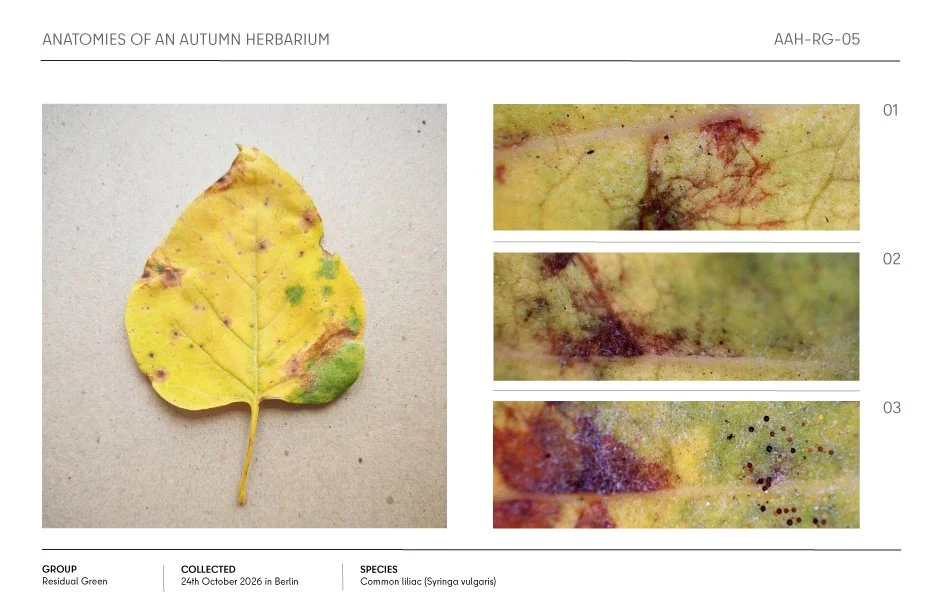
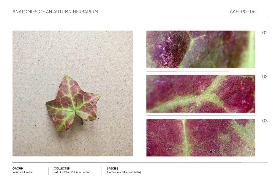
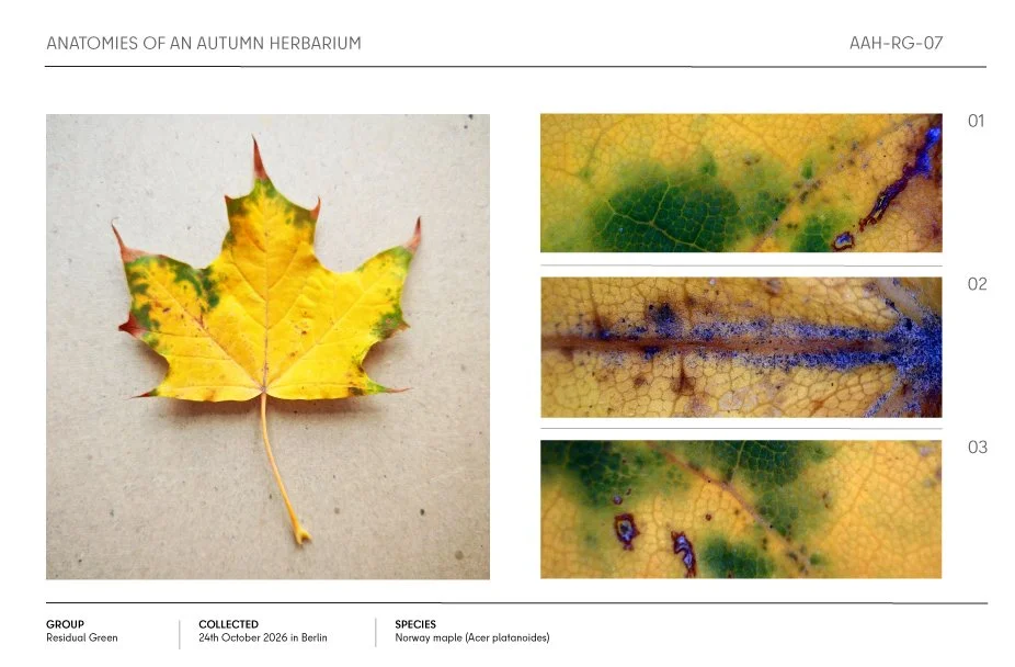
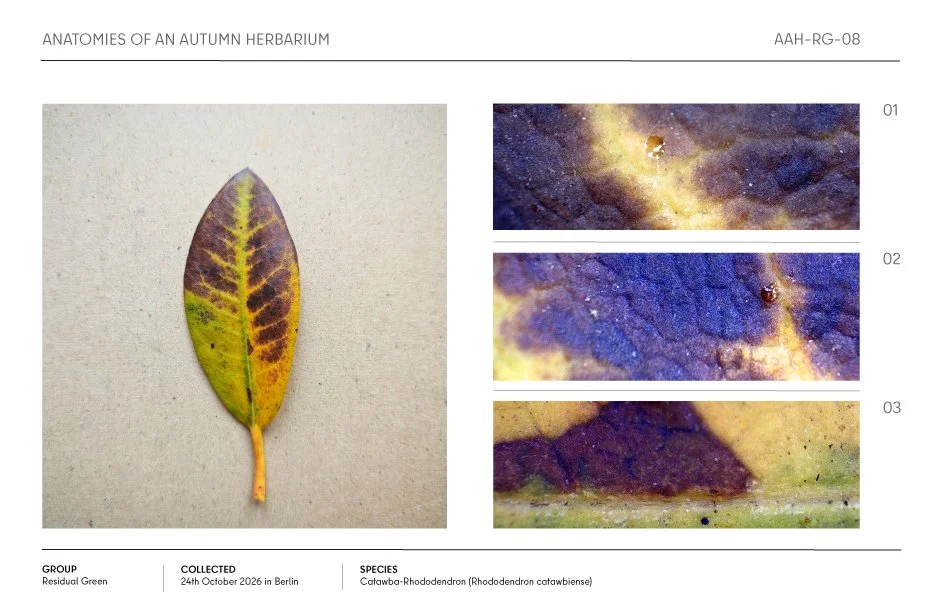
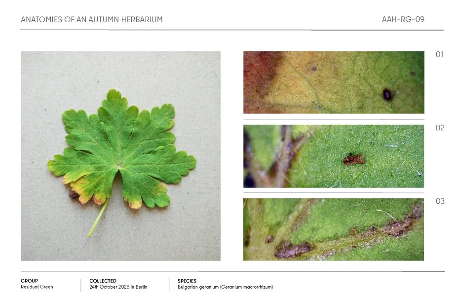
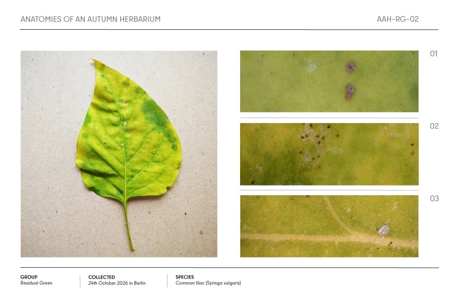
Green islands, narrow margins and vein patterns remain visible within leaves whose surrounding surfaces have turned yellow, red or brown. Their uneven distribution reveals a body undergoing change in different places and to different degrees. Some green areas follow the leaf’s branching architecture, while others appear as isolated patches within a contrasting field. Whole-leaf photographs establish these relationships, and microscopic views bring their boundaries and textures into closer focus. Within the project’s investigation of water withdrawal, these areas become starting points for examining how colour, structure and material condition relate to one another.
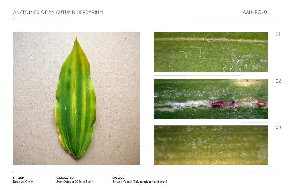
Autumn herbarium sheet showing a green Solomon’s seal leaf with three labeled close-up detail photographs.

Autumn common lilac leaf herbarium sheet with three close-up views of leaf spots and surface details.

Autumn herbarium sheet showing a yellow-green common lilac leaf with three labeled close-up details.

Autumn herbarium specimen of a yellowing common lilac leaf with green and reddish-brown mottling, shown with three close-up detail photos.

Autumn herbarium specimen sheet featuring a yellowing common lilac leaf with green patches and reddish-brown spots, plus three magnified detail views.

Autumn common ivy leaf with green veins and burgundy-red foliage, shown with three close-up texture details in a herbarium specimen layout.

Autumn Norway maple leaf herbarium specimen with three microscopic detail views showing yellow-green foliage, veins, and leaf surface damage

Autumn herbarium page featuring a yellow and purple Catawba rhododendron leaf with three magnified texture details.

Autumn herbarium specimen of a Bulgarian geranium leaf with yellowing edges and three close-up views of leaf surface details